10 an inflamed appendix demonstrates restricted diffusion 10 the appendix may be filled with high t2 fluid or edema which decreases in signal intensity if the fluid is.
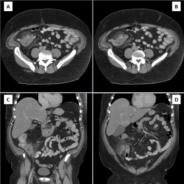
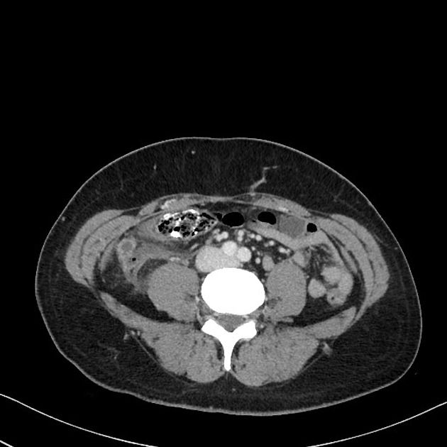
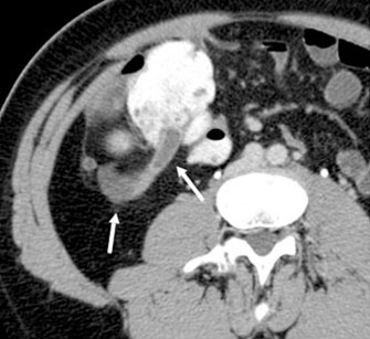
Appendicitis fluid in paracolic gutter.
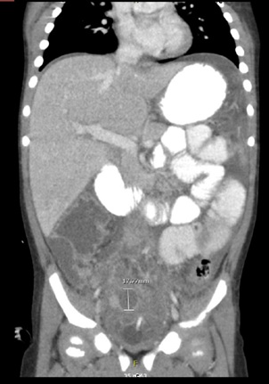
Perforated appendicitis with free fluid.
The patient presented with an elevated white blood cell count and right upper quadrant pain.
Infected peritoneal fluids get a passageway through these gutters to other compartments of the abdominal cavity.
Drainage in patients lying down can be reversed with infectious material from an infected appendix located near the cecum running up the right paracolic gutter.
Return to ultrasound findings.
Fluid from an infected appendix can track up the right paracolic gutter to the hepatorenal recess.
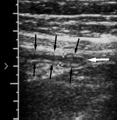
Anterior segment arrow is partially compressed with flattened oval shape and normal diameter of 5 mm but posterior segment arrowhead is rounded with diameter of 7 mm.
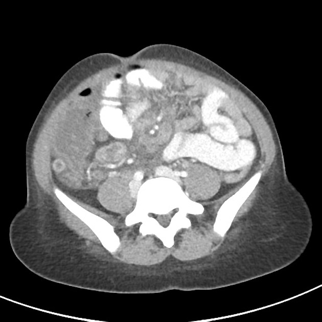
Surgical drains were placed in the pelvis and right paracolic gutter.
Left there is pericholecystic fluid and free fluid in the right paracolic gutter which is caused by.
Dense fluid may suggest hemoperitoneum especially in the context of trauma.
In the abdominal cavity fluid leaches from the colon into either the left or the right lateral gutter and then drains down the gutter into the pelvic area.
Trace periappendiceal hypoechoic fluid is seen which is nonspecific and may relate to edema or trace dependent fluid in paracolic gutter.
These gutters are clinically important because they allow a passage for infectious fluids from different compartments of the abdomen.
The fluid from the infected appendix can track the right paracolic gutter into the hepatorenal recess.
It can be compared to fluid in the gallbladder or stomach.
5 year old girl with a two day history of nausea vomiting fever and abdominal pain presented to the emergency room.
The presence of the gutters also puts the lesser sac at risk of abscission as infected fluids from pancreas and appendix formed due to pancreatitis and appendicitis respectively will accumulate in the left sac.
The findings may be similar to those seen in acute appendicitis including pericaecal inflammatory change abscess formation fluid in the right paracolic gutter cecal wall thickening and an ileocecal mass.
On mri the appearance of acute appendicitis includes an appendiceal diameter 7 mm and adjacent fat stranding that is often best appreciated on t2 fat saturated sequences.


